First Place
 Photographer: Chantelle Venter
Title: Red blood cell hugging a fibrin fibre
The growth in the industrial sector has caused some unfavourable environmental effects. Heavy metals have become synonymous with industrial pollution due to their toxicological and physiological effects on the ecosystem. This is especially true in South Africa, a country that has a thriving industrial sector and not always the means to correctly dispose of toxic materials, like heavy metals, which then enter the air and water supplies. People living in the areas near the polluted air and/or water are the most affected and not only by one metal at a time, but most likely to a combination of metals. This image was taken as part of a PhD study that evaluated the effects of the heavy metals cadmium (Cd) and chromium (Cr) alone and in combination on the coagulation system of Sprague-Dawley rats exposed to the heavy metals alone and in combination over a period of 28 days. This image is a scanning electron micrograph exposed to Cd and Cr in combination and depicts a red blood cell folding itself around the fibrin fibre in a blood clot. It indicates that the red blood cells membrane is altered due to the exposure to the heavy metals and thus easily deforms when a fibrin fibre lays over it.
Second Place
 Photographer: Shivan Parusnath
Title: Honeydew harvest
The Pullen Nature Reserve in Mpumalanga belongs to Wits’ School of Animal, Plant and Environmental Sciences (i.e. Biology), and is used as a research and teaching facility. But being fairly wild and isolated, it is also a fantastic macro photography spot. There, I have been lucky enough to photograph many different organisms, from snakes and frogs to spiders and ants while on teaching fieldtrips, and now get the opportunity to use these photographs as teaching tools – both when teaching photography, and biology.
This is a photo of an ant tending to its aphid colony that I took at the Pullen Reserve during an ecology field trip. I called this photo “Honeydew harvest”, because these ants effectively “farm” colonies of aphids for their honeydew – a sugar-rich sticky liquid secreted from the aphids, which is a valuable food source to the ants. In return, the ants protect their aphid colonies from predators such as ladybugs and wasps. This is known as a mutually symbiotic relationship – and specifically, the positive interspecies association between ants and other organisms is known as myrmecophily.
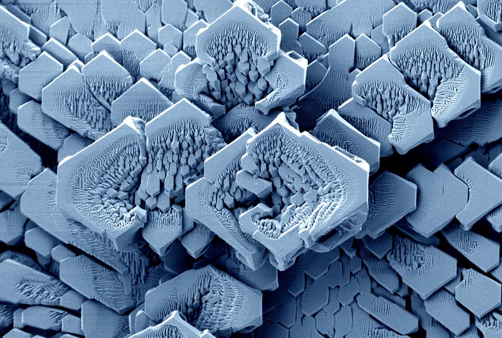 Photographer: Andre Botha
Title: AgS Single crystals
Experimental grown crystals. The growth mechanism of Silver single crystals due to sulfur vapor. The same type of crystals appear on Silver tableware in the form of a purple tarnish due to the atmospheric conditions of every-days air pollution. It will also be visible on any product made from pure silver if you let it in an open environment for a few days.
 Photographer: Tamarin Jurgens
Title: Web of colour
While this image may look like a mass of incredibly colourful spider webs, it is in fact the tubulin microtubule structures of breast cancer cells. Studying breast cancer cells and the mechanisms that are utilised by these cells to migrate throughout the body are incredibly fascinating and vital to the research of metastasis. The cause of many cancer deaths is secondary tumours and not primary tumours. Cytoskeletal proteins like actin (red – stained using phalloidin conjugated to a fluorescent dye) and tubulin (green – stained using primary and secondary fluorescent antibodies) play a huge role in cell migration as well as cell division. DAPI is used to stain the nuclei of the cell (blue) so that each cell, and specifically dividing cells, can be identified. The cell in the top left corner of the image is in the beginning stages of dividing, the actin cytoskeleton is reorganising to form a rounded cell. Confocal microscopy was utilised to obtain this image at a magnification of 63x.
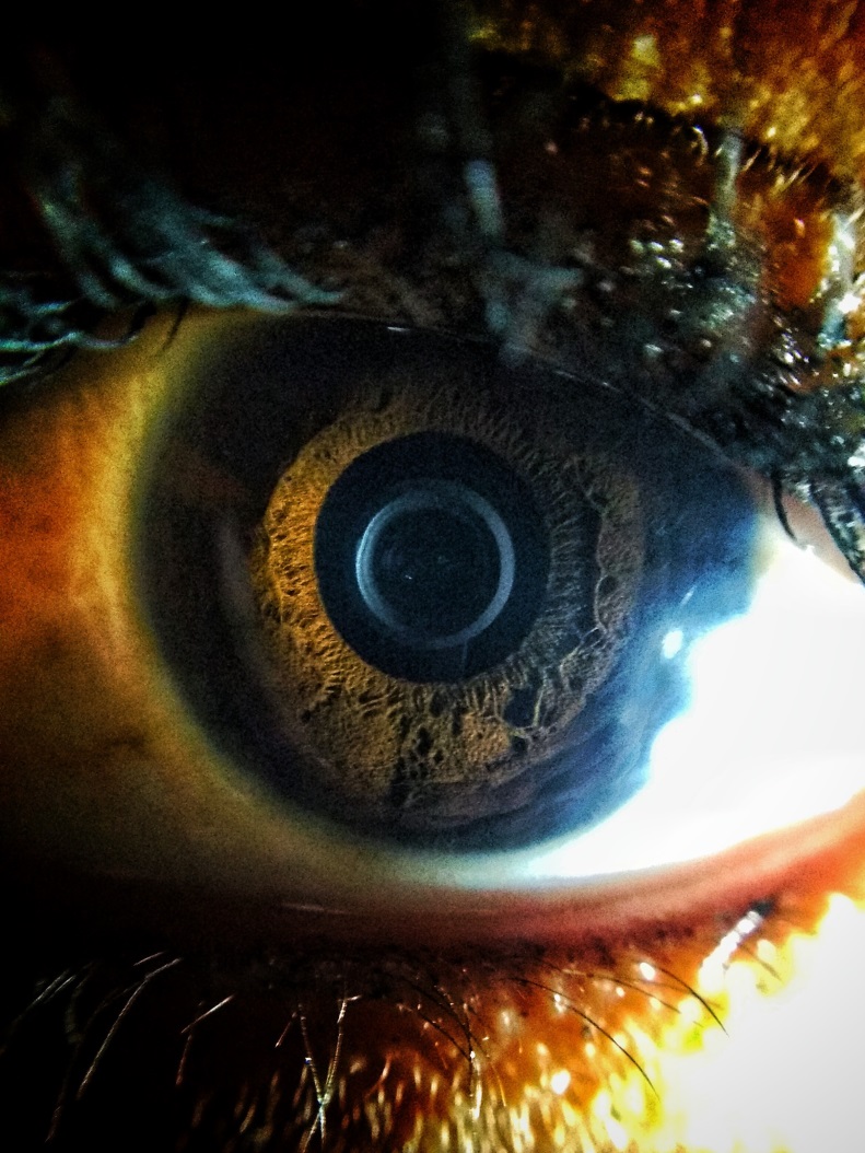 Photographer: Sibusiso Biyela
Title: The naked eye
This is an image of my sister’s eye in close up. I took it with relatively cheap smartphone with a macro lens I bought for less than R20. What this picture means for me as a science communicator is that there is amazing science all around us and it is ironic that in this picture, my sister and I noticed these amazing features in her eye that are not always visible to the “naked” eye.
The animals are then given a stimulative to wake them up, and are safely released in the location of capture.
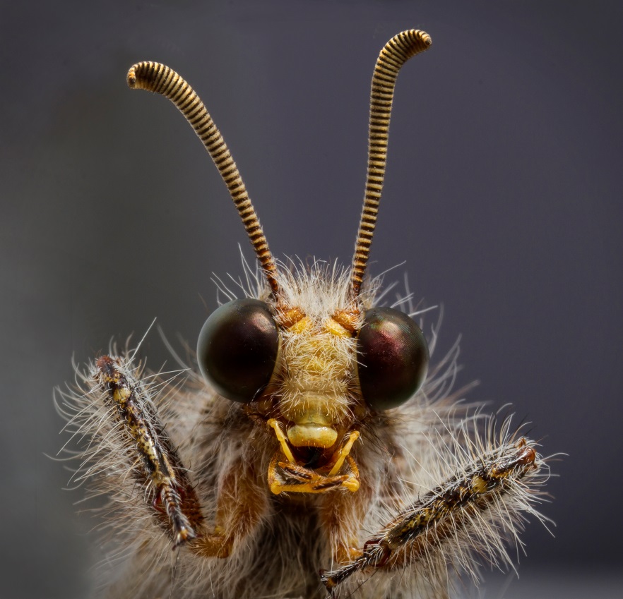 Photographer: Elize Eveleigh
Title: Adult Antlion
The antlion, is neither an ant nor a lion but is from of a group of insects (order Neuroptera) that includes some of Insecta’s weirdest, fiercest and most specialized predators. Their delicate beautiful adult form is a far cry from their fanged larval forms, which trap ants and other small insects in pits dug into the ground. Antlion is a name applied to a group of about 2,000 species of insects in the family Myrmeleontidae. Strictly speaking, the term “antlion” applies to the larval form of the members of this family. What is interesting about certain species like this one is that it demonstrates extreme size difference between the larval and adult body, stretching their mass almost literally to the breaking point with an incredibly thin, fragile adult exoskeleton. Since the larvae only need to back themselves into a hiding place like sand, exposing only their fangs, antlions of all species are incapable of walking forward. Using their flexible posterior to drag themselves backwards as they kick with their legs. Stranger still, they lack an anus, retaining all bodily waste until the pupal stage. Indeed a weird and wonderful creature of nature.
 Photographer: Martin Giger
Title: Surrealism is real
In a surreal conflation of shapes and colours, a peridotite reveals its hidden beauty in a cross-polarised microscopic image. While the interlobate grain shapes are generally taken as proof for crystal growth under equilibrium conditions, the rock has been intensely fractured since its origination. The colours are indeed no “true” colours as seen with the naked eye. Polarised light traversing a crystal may experience a slight twist in orientation, a shift from the direction superimposed on it by the polarising filter. Applying a second filter exactly perpendicular to the first one effectively filters out all light that has not been modified by refraction on the crystal face, now showing every single crystal in its distinct interference colour. Dull, perfectly achromatic rocks may turn into colourful kaleidoscopes of every shade of the rainbow – only against a black background, rendering them luminescent beyond anything. Inclined fractures in the grains accentuate the interference of the crystal faces with the light it is traversed by, turning pale turquoise into a luscious purple.
Click here to read more about the competition.
*The copyright of the photographs entered into the competition remains with the photographer. The photographs may be published in connection with the SA Science Lens competition, acknowledging SAASTA, the SA Science Lens competition and the photographer. For enquiries regarding permission to use photos, please contact Joanne Riley, Science Editor at SAASTA, at joanne@saasta.ac.za or 012 392 9349.
 Joanne Riley
 The South Africa Agency for Science and Technology Advancement (SAASTA) is a business unit of the