Winner
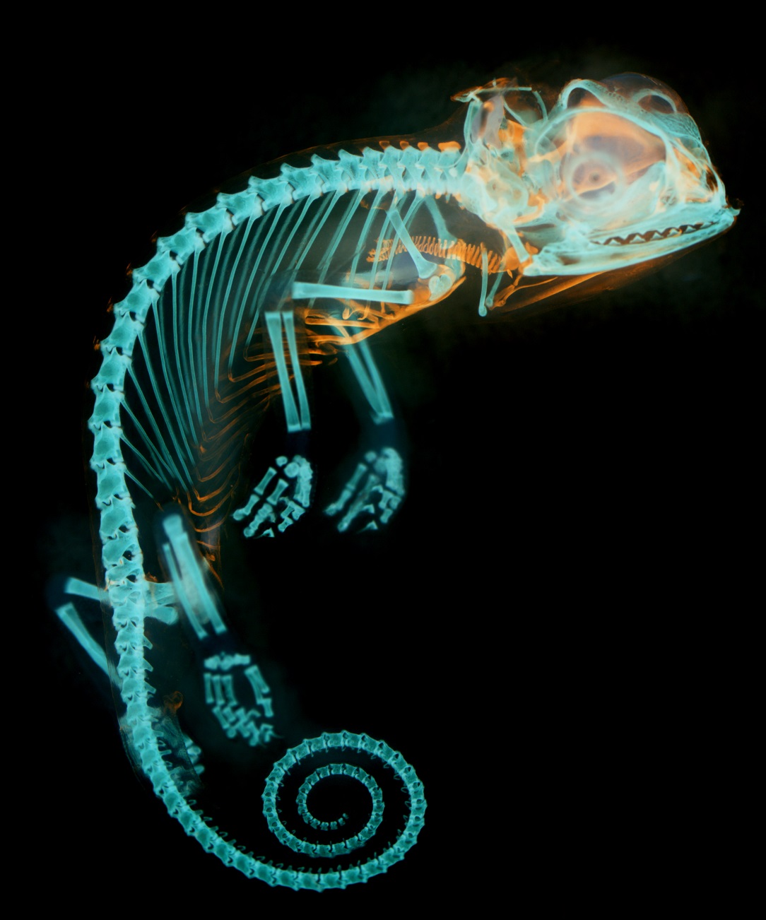
Photographer: Dr Dorit Hockman
Title: Inside Out
A late stage embryo of the species Chamaeleo calyptratus, the veiled chameleon. I removed the skin and used skeletal stains to dye the cartilage and bones of the embryo before using chemical agents to clear the surrounding tissue. I have inverted the image to create an x-ray like quality, which emphasises the developing bones in blue and the cartilage in orange. As the skeleton forms cartilage is gradually replaced by bone. At this stage of development, most of the bones have formed, while the front of the ribs are still formed from cartilage. The cartilage rings that support the oesophagus are also clearly visible. I was given the opportunity to work with these and embryos of a variety of animals as part of the Embryology Course at the Marine Biological Laboratory in Woods Hole, Massachusetts, while doing my PhD at the University of Cambridge. By comparing the processes of embryonic development, such as the rate of skeleton growth, across different animals we can gain insight into how evolution acts on these processes to allow the diversity of form in the animal kingdom.
Runner Up
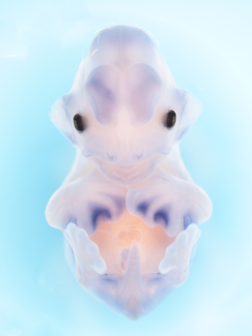
Photographer: Mandy Mason
Title: What’s in a Wing?
This image shows an embryonic bat (Miniopterus natalensis) at a stage of development where the hands form distinctive elongated digits and interdigtital webbing of the wings. The open mouth of the embryo and flexed forelimbs that are tucked beneath its chin are characteristic of this stage (Carollia stage 17), which is commonly known as ‘Tongue Out’. The bat’s hindlimbs, which form short free digits, project outwards. This embryo has undergone an in situ hybridisation experiment, whereby a gene’s activity is visualised through the staining (in purple) of the mRNA molecule that it transcribes. In this particular experiment, the gene Meis2, shows unique expressions in the developing tissues that will form the membranes (chiropatagia between the digits, plagiopatagia between the forelimb and hindlimb and the uropatagia between the legs and the tail). This points towards a role for this gene in the evolution and formation of these structures. The evolution and development of the bat wing remains enigmatic and provides a remarkable contrast to embryonic studies of the conventional model organisms.
Special Mentions
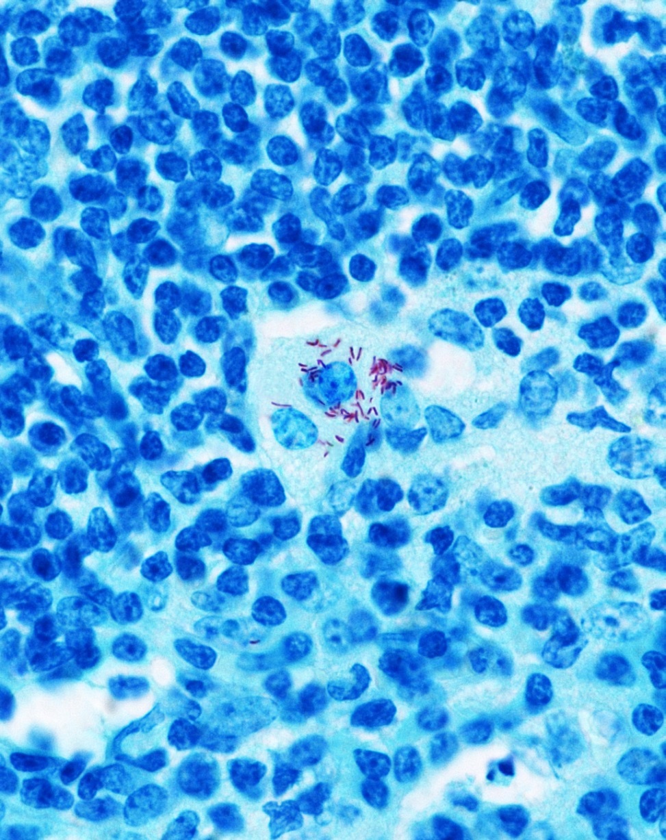
Photographer: Jennifer Hoving
Title: Mycobacterium Tuberculosis in Living Cells
With the high TB infection rate in South Africa we would benefit substantially from new diagnostic tools and treatment strategies. For this reason, our research group within the Institute of Infectious Disease and Molecular Medicine at the University of Cape Town focuses on host immune response to disease with a specific interest in TB and AIDS related fungal infection. This image shows the pathogenic bacteria Mycobacterium tuberculosis (pink) infecting the cells of an inflamed mouse lung (blue). This image was taken in a gene deficient (CLECSF8) mouse that was susceptible to the bacteria and helped us identify a key receptor involved in the control of tuberculosis. We showed for the first time that a pattern recognition receptor is important in controlling a clinical strain of M. Tuberculosis, as removing the receptor resulted in 20% death of the mice. This research tool could be used in the future to help identify those most likely to get the infection and was published in Cell Host and Microbe earlier this year (Wilson, Marakalala, Hoving, van Laarhoven et al CHM 2015).
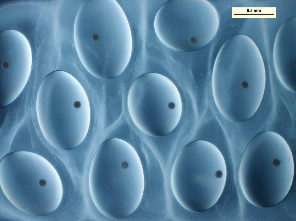
Photographer: James Welsey-Smith
Title: Snail Eggs
This image shows eggs of the snail Lymnaea spp., and the work was part of a Ph.D. study aimed at characterising this aquatic species. The low contrast of this unstained living sample was overcome by using Differential Interference Optics. The relatively thick nature of the sample was beyond the depth of focus of the objective used. In order to obtain a well-focused image, approximately 10 through-focus images were captured, aligned and stacked, rendering the necessary extended depth of field.
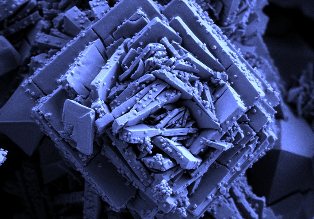
Photographer: Sharon Eggers
Title: Crystal Rose
This Image shows the crystal structure of a MOF (Metal Organis Framework)compound. It is important to be able to see the size and shapes of the crystals which influence the properties of the material.
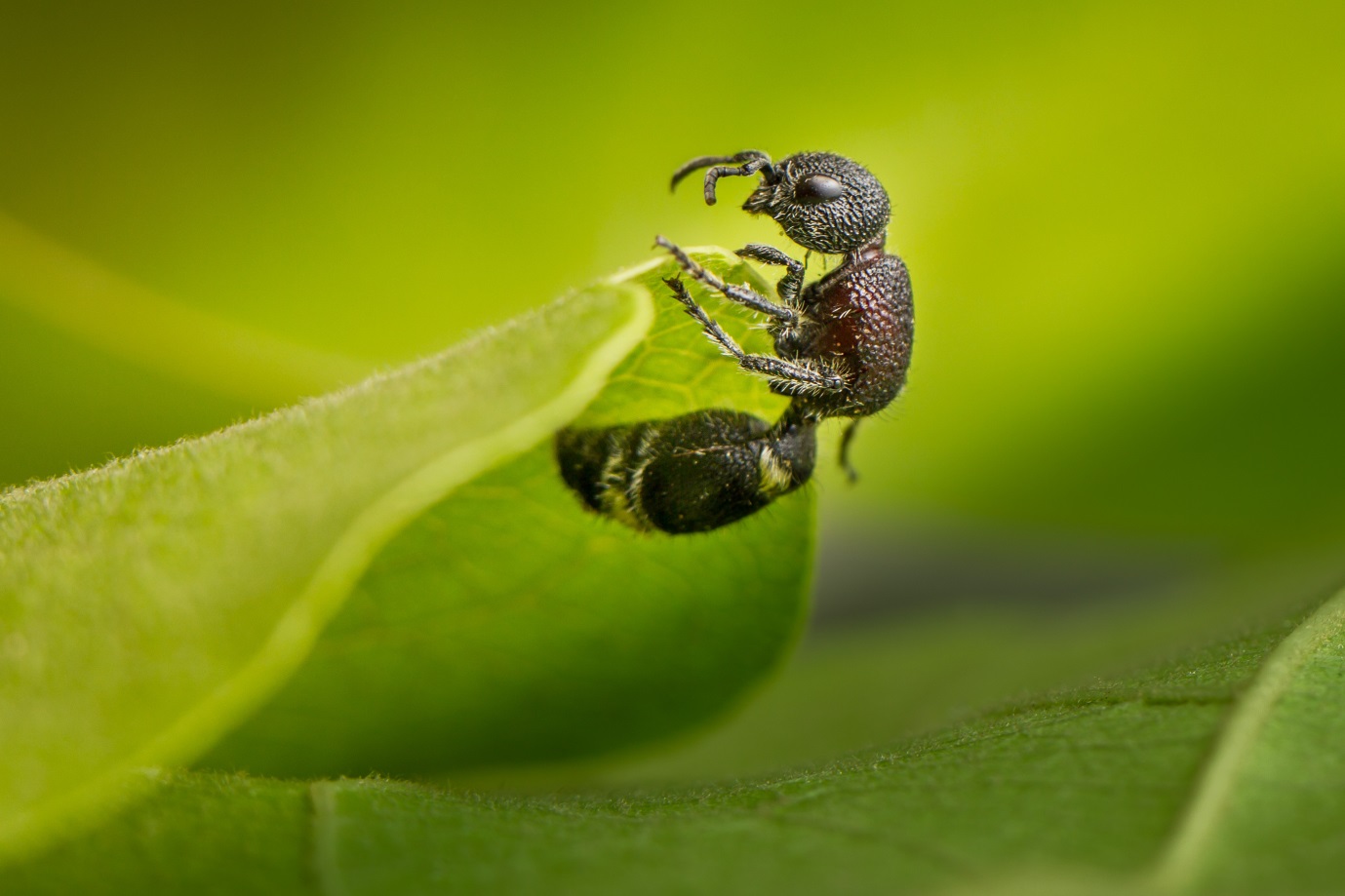
Photographer: Morgan Trimble
Title: Lucky Velvet Ant
I came across this critter in Majete Wildlife Reserve in Malawi. Majete is in the process of being restored after losing several of its mammal species to local extinction due to human encroachment in the park and poor enforcement of environmental laws. Though many mammal species have been reintroduced, many of the smaller wildlife species such as this velvet ant, likely clung on in the park. Park guide, Jimmy, told me that this velvet ant is considered a good luck charm in Malawi. Unluckily for the velvet ant, they are also sometimes used in traditional medicine. Interestingly the velvet ant is not an ant at all, but a type of flightless wasp species in the Mutillidae family. “Velvet” in their common name refers to the dense pile of hair on their bodies, visible in this macro shot. Like wasps, they can deliver a powerful sting. The flightless females, like this one, look a lot like ants, but the males have wings.
Copyright Notice
These SA Science Lens images are protected by copyright. Should anyone wish to use these images, please contact SAASTA at sciencelens@saasta.ac.za.
 The South Africa Agency for Science and Technology Advancement (SAASTA) is a business unit of the